
In our series of awesome MRI scanners …

By Biomedicine in museums

… check out these pics from the installation of the brand new 32 tons heavy 7 Tesla experimental whole-body MRI at the Charité Hospital in Berlin (via Medgadget). Note the caption to the third image: the only thing the blogpost-writer knows about the guy inside the magnet is that he didn’t wear his nipple rings that particular day 🙂

These super-machines support the ‘contemporary-medical-museum doctrine’, which I presented to a conference group on contemporary medical museums in the Boerhaave Museum in Leiden some years ago (and which some of my colleagues thought was baloney). On the one hand significant medical objects are becoming smaller and smaller (nanomedicine), on the other hand some instruments are becoming bigger and bigger (like these scanners). In contrast, human-sized medical artefacts are becoming less and less signifiant for what’s going on in the medical system.

This tendency — which is analogous to the situation in particle physics where bigger and bigger accelerators are needed to provide insights into progressively more tiny scientific objects — makes medical museum acquisitioning and collection management increasingly difficult. Today medical museum storage rooms are filled with human sized artefacts. But the storage rooms of the future will probably be more like a combination of those they have in museums of transportation (with really big objects like airplanes) and those they have in biobanks (liquid nitrogen freezers to keep molecular and cellular samples).

In other words the major future problem for medical museums is a peculiar combination of lack of space and lack of visibility.

Biomedical autobiographies

By Biomedicine in museums

Having an affection for scientific biography/autobiography, I was thinking of how I could possibly engage with the conference ‘Academic Autobiography, Intellectual History, and Cultural Memory in the 20th Century’ to be held 26-28 March, 2009 at Universidad de Navarra in Spain.

The aim of the meeting is to engage with current discussions among historians, literary critics, anthropologists, sociologists, etc. about how intellectual history and cultural memory may be developed, articulated, and promoted through autobiography. In other words, the organizers emphasise themes like

  • the academic as author/historian
  • academic life writing as history or cultural discourse
  • academic autobiography as intellectual history
  • life writing and the definitions of academic disciplines
  • the intersection between private and public lives in academic autobiographies
  • academic autobiography as a literary or historical genre
  • the ways in which the notion of literary or historical discourse may be rethought in the context of this form of writing
  • the ways academic autobiographies challenge our notions of historiography or literary analysis.

The limitaton to the 20th century is fine. But what about the organizers’ understanding of what counts as ‘academic autobiography’? Humanities and social science scholars like historian Eric Hobsbawm, anthropologist Clifford Geertz, cultural critic Edward Said and others are mentioned, but scientists are apparenty not thought of as ‘academics’ in this context.

Yet the genre of scientific autobiography has a long and interesting history (also in the 20th century), which could be drawn upon for qualifying the discussion. Just think about how James D. Watson’s first autobiography, The Double Helix (1968) has changed contemporary perceptions of the life sciences! Its cultural impact led to it being ranked among the top ten titles on Modern Library’s 1998 list of best 20th century non-fiction books together with classics like Rachel Carson’s Silent Spring and John Maynard Keynes’ General Theory of Employment, Interest and Money. (Watson’s second autobiography, Avoid Boring People [2007; review here] hasn’t made it to any list yet, though).

How would studies of scientific autobiography add to the conference theme? In many ways scientific autobiography is not much different from literary or political autobiography. Except in one crucial way, namely that the author’s understanding of self in terms of science may influence his/her understanding of what it means to write an autobiography.

Biotech maverick Craig Venter recently employed this autobiographical trope in A Life Decoded (review here). He claimed that he saw his life narrative as the result of a genome that writes reflexively about itself. It wasn’t very convincingly done, and at first I dismissed the idea as terribly naïve. But thinking about it again, I believe he points to an interesting space for future self-writing.

As the postgenomic worldview — and especially the results of molecular neuroscience — is spreading in our culture (at least the secular part of it; religiously based cultures are probably immune), more and more people will probably understand their selves as complex biological systems, as intricate protein-protein interaction and metabolic machines. To think about oneself as a bundle of biomolecular reactions may, I suggest, become a pervasive existential motif in future autobiographical narratives. Michel Houellebecq has already played with similar ideas in The Possibility of an Island (see here).

I don’t think it’s as far-fetched as it immediately sounds. Freudian understandings of the self became an immensely influential trope in biographical and autobiographical writing in the first half of the 20th century. The explanatory power of postgenomic, molecular neuroscience may become equally influentual for the understanding of self in autobiographical writing. Venter’s attempt wasn’t convincing, but other and better attempts will hopefully follow.

Anyway, I probably won’t have time to send something in — they want a 500-word abstract before 15 October. More info about the conference here.

Video publications will be indexed in MEDLINE/Pubmed

By Biomedicine in museums

Back in 2006 we wrote enthusiastically about the first issue of the online Journal of Visualized Experiments — the aim of which is to publish video films of experimental work to help apply laboratory protocols. A “YouTube for test tubes”, as it was then called.

Since then JoVE has published more than 200 videos of laboratory procedures. Now (says Nature, 4 Sept, p 13), the content of JoVE will be indexed in the MEDLINE base and thus available through the PubMed search engine. An interesting policy move from the side of the National Library of Medicine, because it means that the video format is now being endorsed on a par with text articles as an acknowledged form of publication (cf. how NLM last year began to endorse blogs as publications, see earlier post here).

JoVE is a potentially great source for exhibitions on contemporary biomedicine, either for direct use, or indirectly, as an inspiration for producing new videos for public outreach of laboratory practices. The videos demonstrate how the laboratory has its roots in manual labour, and are a reminder about how thoroughly materially grounded biomedical practices are.

These and similar video repositories are a great complement to ethnographic description of laboratory practice and may contribute further to the rejuvenation of studies of the laboratory as a knowledge production space that Dominique Vinck and others are currently involved in (see, for example the special issue on laboratory studies in Revue d’anthropologie des connaissances, vol. 1 (n° 2), 2007).

There are curators — and then there are biocurators

By Biomedicine in museums

When I hear the word ‘curator’ I usually think of museums. Museum curators are the sort of knowledge workers that take care of stuff in collections and make exhibition narratives possible.

Now I’ve just learned about another — different yet similar — kind of curators. The background is the current enormous growth in the generation of data from life science research. Large-scale sequencing, high-throughput gene-expression analyses, and mass-spectroscopy projects produce huge amounts of gene and protein sequences and other bio-data.

It doesn’t take much thinking to realise that the usefulness of all this data-generating research depends on the scientific community’s ability to make sense of the massive amounts of raw data scattered among millions of scientific papers and hundreds of data bases.

Here’s where the new kind of curators come into the picture. A recent article in Nature (Howe et al., ‘Big data: the future of biocuration’, 4 September) discusses the emergence of “a growing cadre of biologists — ‘biocurators’ — who manage raw biological data, extract information from published literature, develop structured vocabularies to tag data and make the information available online”.

When I read the article it struck me that there are some interesting similarities between museums curators and biocurators. A central aspect of data-curating is annotation — so far, however, says the Nature article, annotation efforts have been limited by a perceived lack of incentive to do the job: “A mechanism tied to career or research advancement may be required before community curation can be established as a broadly accepted and productive scientific endeavour.”

In other words, what is needed to speed up bio-curation is “improvement in academic reputation or impact, career advancement and better funding chances”. Further, academic departments and funding agencies “should consider community annotation as a productive contribution to the scientific research corpus and a natural extension of the publication process”.

Sounds like a parallell to the situation among museum curators. They too are working in the shadow of university scholars. Curatorial work in museums doesn’t have the same reputation as university research. So whether you work as a museum curator or as a bio-curator, you risk being invisible among ‘real’ researchers/scholars and suffering a lack of scholarly status.

Wonder if this has something to do with the fact that curatorial work is ‘care work’ (from Latin ‘cura’ = care)?

Read more in Nature’s special issue on ‘Big Data’ here.

Biotech exhibitions between fascination / fetischism and resignation / hostility

By Biomedicine in museums

The Lentos Art Museum in Linz, Austria, has just opened an exhibition called ‘Ecology of Techno Mind’ curated by Slovenian “art-is-the-evil-of-culture” curator Jurij Krpan (thanks to Ingeborg Reichle for the tip). It contains, among other things, an array of art works relating to biotechnology and computerized medicine made in order to “eine Welt im Wandel zu verstehen” [to understand a changing world].

What’s interesting from a medical museum point of view is that the Linz exhibition (according to the website, I haven’t seen it IRL) tries to navigate between “die Total-Faszination von Wissenschaft und Technik” [the total fascination with science and technology] and the “Resignation und Feindseligkeit gegenüber Technik” [resignation and hostility toward technology]; it’s dealing head-on with biotech and medicine but not from the angle of “Fetischismus” or “Romantisieren von Interfaces und elektronischem Kabelsalat”. Sounds like a familiar problem for science, technology and medical museum curators :-). Maybe we could learn something from how Krpan, Director of the Kapelica Gallery in Ljubljana, handles this sensitive issue.

Transforming dead bodies into scientific and artistic objects

By Biomedicine in museums

Tomorrow, Tuesday 9 September at noon, we’re having a lunch seminar with Sebastian Abrahamsson, doctoral student at Jesus College, University of Oxford. Sebastian will speak about ‘Dead bodies in science and art’ — a topic which is very central to our research and exhibition work here at Medical Museion, so we’re expecting a lively discussion.

Here’s Sebastian’s abstract:

My research is interested in the ways in which dead human bodies are transformed into artistic and/or scientific objects.

As a point of departure, my project maps the work of an artist (www.angelaspalmer.com) who – together with museum curators, radiologists and Egyptologists – has been working with a mummy child making his body and journey the focus of an art exhibition in a gallery in London. This work brings this particular body through a set of heterogeneous milieus (museum, hospital, studio, and gallery) and environments – each with its own practices, routines and experts – thus changing the significance and interpretation of the body at each stop that it makes: going from museum object to aesthetizised sculptures.

Secondly, I look at the changing status of dead bodies (educational and scientific specimen turning into contested and controversial human remains) in British museums, taking Manchester Museum and their Egyptian collection as an example. Recently three of their unwrapped mummies were covered, provoking massive public reactions (mainly negative). The museum curators had clearly taken a controversial decision, but why this outrage? Here I argue the mummies become the focal point of a sort of public experiment, the outcome of which is still to be decided.

As a final example my project looks at Von Hagens’ Body Worlds as the site where a scientific/educational and artistic ethos come together to stage an encounter with dead bodies, where the above distinction between art and science is more or less erased. Out of these three examples emerges the question concerning how we can experience encounters with dead bodies differently.

If you want to attend, please call our technician Folke Jørgensen at +45 3532 3822, who can give you more detailed info about the location of the seminar room in Fredericiagade 18, Copenhagen, and how to get into the house. Bring your lunch! 

(Image above from Angela Palmer’s website)

Museum exhibitions as products and generators of scholarship

By Biomedicine in museums

Just a few words about the upcoming conference ‘The Exhibition as Product and Generator of Scholarship’ at Deutsches Museum in Munich, 27-28 November 2008 — a follow-up on the conference Research and Museums that was held in Stockholm in May last year.

It will be an interesting conference because — by bringing together exhibition makers, museum experts, designers, artists, cultural studies scholars and historians of science and technology — it addresses the core issue in our work here at Medical Museion, namely, the interplay between research and museum work. Two of us (Martha and myself) will present papers.

The motto of the meeting is: “No exhibition without scholarship”. In other words: museum exhibitions aren’t just about visualizing results of historical and other kinds of museum-relevant research results; they also stimulate academic scholarship and generate new research question and new knowledge:

How can researchers take advantage of this opportunity? In which way can scholarly arguments be translated into spatial arrangement and at the same time kept serviceable for reading and citing by later recipients? What might the results of the scholarly examination of an exhibition look like? Unlike for printed texts, the traditional publication media of scholarship, common standards of terminology and argumentation for exhibitions have yet to emerge. What exactly is the role of the objects on display? Recent history of science and technology has intensively interrogated the epistemic quality of these material sources of research. Yet how do the objects unfold their properties in being staged for exhibition purposes?

Sessions:

1) What is this thing called exhibition? Reflections on object, text and space

  • Ulrich Raulff (Marbach), Old answers, new questions: What do exhibitions really produce?
  • Jochen Brüning (Berlin), Exhibitions vs. publications. On scientific achievements and their evaluation
  • Martha Fleming (Copenhagen/Toronto), Thinking through objects
  • Commentary: Lorraine Daston (Berlin)

2) Stories on display. What and how do we see in exhibitions?

  • Uwe W. Brückner (Stuttgart), Scenography – opera as model for integrative design
  • Stefan Iglhaut (Berlin), Story telling and scenography: Strategies of science communication in exhibitions
  • Commentary: Anke te Heesen (Tübingen)

3) History of science, objects, exhibitions: Interrelations, transitions, transformations

  • Hans-Jörg Rheinberger (Berlin), Making visible. Visualization in the sciences – and in exhibitions?
  • Ulrich Großmann (Nuremberg), The Challenge of Objects – CIHA Congress 2012. The object in the focus of arthistorical studies
  • Thomas Söderqvist (Copenhagen), Do things talk?
  • Commentary: Falk Müller (Frankfurt)

4) More than history of science?! Exhibitions, research, and the public

  • Mosbrugger (Frankfurt/M.), Natural history research and exhibitions – a hermeneutical cycle
  • Robert Bud (London), Power, belief and trust: a context for scholarly priorities in the history of science
  • Ad Maas (Leiden), Tearing down the altar. A new view of displaying scientific intruments in Museum Boerhaave
  • Commentary: Jochen Hennig (Berlin)

5) Making exhibitions: Concepts, constraints, critique

  • Jürgen Renn (Berlin), Exhibitions as history of science in action
  • Walter Hauser (Munich), Artefacts, visuals and topography as evidence: Working on an exhibition on nano- and biotechnology
  • Thomas Schnalke (Berlin), Arguing with objects. The exhibition as a scientific format of publication
  • Commentary: Karsten Gaulke (Kassel)

The conference is organised for the Max Planck Research Network ‘The History of Scientific Objects’ by Helmuth Trischler, Christian Sichau and Susanne Pickert at Deutsches Museum. You are welcome to contact Susanne Pickert at s.pickert@deutsches-museum.de if you want to attend.

Biomedical images online for exhibition purposes

By Biomedicine in museums

There are many ways of finding biomedical images on the web for exhibition use, and some are better the others.

Getty Images, which is otherwise a fantastic online repository of professional high-quality images, is practically useless for a small museum like ours. Search ‘protein’, for example, and you get over a thousand images of eggs, tofu and pork meat and other everyday stuff associated with proteins, but very few scientific images — and the few they have are excruciatingly expensive (the cost for using a single ‘beautiful’ electrophoresis image is around 600 USD). I dare not reproduce even a thumbnail size image from Getty Images here because we risk being persued for violating their intellectual property rights; in fact, I’m not even allowed to show their company logo!).

Wellcome Images is much more useful for our exhibition purposes. Search ‘protein’ and you get over 500 images of protein molecule models, fancy microscopic images, high-tech protein research instruments, and so forth — like this image of a computer-enhanced analysis of a 2D protein gel (credit: Nicoletta Baloyianni; Wellcome Images). They’ve also got some good images of protein-rich food items and other everyday stuff. And best of all for a poor university museum like ours — their images are freely available for download under a Creative Commons licence.

Both Getty Images and Wellcome Images are collections of ‘beautiful images’ — that is, the kind of immediately aesthetically pleasing pics that science magazines fill their pages with and scientists like to hang on their office doors. The Yale Image Finder search engine developed by Michael Krauthammer’s lab at Yale Center for Medical Informatics is different (read more about the project here). It allows you to find the image content of (presently) some 35,000 open access articles from PubMed Central by key word searches in figure texts, captions, abstracts, titles and even full article texts. For example, searching for ‘protein’ in captions and figure texts gave 19,000 hits, like the Western blog analysis image to the right.

Yale Image Finder is developed as a tool for scientists, not for curators. Right now it’s somewhat bothersome to use, because you have to sift through so much material. But it gives you rapid access to hundreds of thousands of close-to-the bench kind of images which do not find their way to Wellcome Images, and as such it may become a useful supplement to the ‘beautiful image’ online repositories.

Ideas for a home-made pathological museum

By Biomedicine in museums

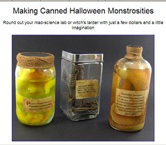
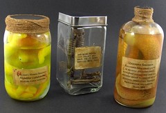
Ever thought about building your own collection of medical wet specimens? Spending your evenings and gloomy sleepless nights in the garage putting your family’s and friends’ pickled organs and body parts in jars? Founding a clandestine horror show?

Well, it’s not for real. Yet. It’s another Halloween idea:


(from I Make Projects.com; thanks to Paul at NMHM for the tip)

Physics meets biology: Perspectives from philosophy, history and science (Edinburgh, 18-20 November)

By Biomedicine in museums

Have forgotten to announce the ‘Physics meets biology’ meeting at the Royal Society of Edinburgh, 18-20 November 2008. Interesting because, as the organisers say, collaboration between physicists and biologists has generated a host of fascinating philosophical problems; e.g., they often disagree about the role of hypothesis in research, what an explanation is, etc. Such differences have practical consequences for interdisciplinary research and also for the border area between applied physics and biomedicine. The Edinburgh meeting brings together physicists, biologists, historians, philosophers and science policy makers; confirmed speakers include Evelyn Fox Keller (Science and Technology Studies, MIT), Kevin Dunbar (Psychology/Brain Science & Education, Dartmouth), Steven French (History and Philosophy of Science, Leeds), Michel Morange (Biology & History and Philosophy of Science and Technology, École Normale Supérieure, Paris), Gregory Radick (History and Philosophy of Science, Leeds), Otávio Bueno (Philosophy, University of Miami), and Darrell Rowbottom (Philosophy, University of Bristol). More info on http://www.ph.ed.ac.uk/pmb2008.