Monthly Archives

July 2009

Use the current lingua franca, please

By Biomedicine in museums

Two months ago I praised John Harley Warner’s and Jim Edmonson’s book Dissection: Photographs of a Rite of Passage in America, 1880-1930.

As Kirsten Jungersen points out in a comment, one of our former staff members here at Medical Museion, Mikkel Jessen, wrote about dissection as a rite-of-passage in an article in the journal Bibliotek for Læger already in September 2002 (pp. 260-69).

Mikkel’s is a short but excellent article on four different ways in which dissection has been displayed: Rembrandt’s ‘De anatomishe les’, Hogarth’s ‘The four stages of cruelty’, Simonet’s ‘La autopsia’, and a photo of a staged dissection at the Royal Academy of Surgeons in Copenhagen, where the medical students are trained in ‘the necessary kind of inhumanity’.

What triggered this post, however, is that Mikkel’s article is yet another example of how the work of young scholars in small countries remain largely unread outside the small national circle (Bibliotek for Læger publishes in Danish only). Had Mikkel written his piece in English it would have been recognized several years before John and Jim published their excellent book. I mean, he could have been recruited as a PhD-student at Yale, where John works, or whatever.

So Mikkel’s article reminds me how many good opportunities are lost because too many young Danish (Swedish, Norwegian, Estonian etc.) scholars restrict themselves to writing in their mothertongues. Use the current lingua franca, please!

Science exhibitions: curation, design and communication

By Biomedicine in museums

Anastasia Filippoupoliti at the Democritus University of Thrace, Greece
(afilipp@gmail.com) and Graeme Farnell at MuseumsEtc, UK (graeme@museumsetc.com) are soliciting papers for a forthcoming book that will explore:

  • the processes involved in developing new science exhibitions in and for museums
  • the issues involved in transforming scientific ideas or events into exhibitions
  • the challenges faced by museums in communicating science to a wide audience.

Much has been written about the difficulties of disseminating science to the public through a variety of new and traditional media. It is, indeed, a complex subject to tackle in the exhibition space, yet a challenging and multidimensional one.

How best to understand the process of working from scientific data to the ideas-based exhibition? What exactly is lost during the transformation of factual information into an exhibition environment? And more importantly, how can the exhibition work most effectively as a tool for narrating science, its past and present?

They welcome a range of submissions including, but not limited to, the following issues/themes:

  • both theoretical perspectives and case studies relating to science exhibitions
  • exhibition design for science: problems and opportunities
  • successful design techniques and approaches in relation to science displays
  • science communication in the museum: interpretation issues
  • learning activities and science collections
  • developing learning resources for science exhibitions
  • object stories and science learning
  • exhibitions interpreting the history of science

Please submit an abstract (up to 400 words) and a biographical note (up to 250 words) by email to both editors above. Deadline for abstracts and bio 30 September 2009. Selection for inclusion 30 October 2009.

postdoc/PhD position: Communicative barriers between biomedical research and everyday health care in a museum context

By Biomedicine in museums

Medical Museion, University of Copenhagen, invites applications for a 3-year PhD/2-year postdoc position in the projekt ‘Communicative barriers between biomedical research and everyday health care in a museum context’:

The increasingly important role of biomedical knowledge and advanced medical technologies in Western health systems is a challenge to the public communication of and engagement with medicine, especially in science, technology and medical museums. This project aims to develop the understanding of the means through which esoteric biomedical and medico-technological knowledge and practices can be communicated to lay people in a museum exhibition context, with special emphasis on the use of material and visual cultural practices. The project is expected to help construct new physical and web-based exhibition and display practices for science, technology and medical museums.

Medical Museion is an integrated research and museum institution that focuses on the public engagement with the contemporary biomedical sciences. We are especially interested in the interface between biomedicine, material and visual studies, museum studies and studies of the participatory web. See www.corporeality.net/museion.

The salary level for a PhD-candidate in Denmark is 295,000 DKK (approx. € 39,600/ £ 34,000) p.a.

The salary levels for a postdoc is 370,000 DKK (approx. € 50,000 / £ 42,000) p.a. upwards (depending on earlier experience).

The position is part of a larger cross-faculty research program ‘Dissemination and Innovation: Health in Everyday Life’, which in turn is part of a newly established Center for Healthy Aging at the University of Copenhagen (see http://healthyaging.ku.dk).

Applications for the postdoc level shall include CV and publication list; short description of background and motivation for choosing this research project; project description (max. 5 pages); and copies of degrees.

Application for the PhD level shall include a CV; short project proposal (max. 2 pages); academic transcripts; and copies of degrees.

Deadline is 1 September, 2009 at 14.00. Applications must be submitted electronically and in print (4 copies) to:

Assoc. prof. Lene Otto
Section of Ethnology, the SAXO Institute
Faculty of Humanities, Njalsgade 80
DK-2300 Copenhagen, Denmark
lotto@hum.ku.dk

Further information can be obtained from Professor Thomas Söderqvist, ths@sund.ku.dk, or +45 2875 3801.

For the full text of the announcement in context, see http://healthyaging.ku.dk/vacancies.

Why are hospitals associated with the colour green?

By Biomedicine in museums

Ever wondered why hospitals are associated with the colour green? Green surgery scrubs, green operating theatres, green-painted instruments, and so on and so forth.

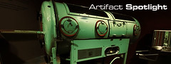
A temporary exhibition called ‘Artifact Spotlight: The Colour of Medicine’ at the Canada Science and Technology Museum in Ottawa shows how the colour green conquered the hospital world during the 20th century:

Green was a popular choice. Surgeons first added “spinach-leaf green” to their clothing in 1914 to reduce glare from traditional hospital whites. In the 1930s, hospital decorators used green to influence patient moods. It carried associations with nature, growth and recovery. Tiled surgical suites, patient rooms, clothing and instruments all went green in the post World War Two era.

The exhibition curator, David Pantalony, is currently exploring the history of the colour green in medical instruments in the period 1950 to 1975 and in medicine in general. Look out for his forthcoming article in the Canadian Medical Association Journal this summer.

Here’s an another image of the exhibition:

Udstilling set fra et designprofessionel udgangspunkt

By Biomedicine in museums

Exponent — et dansk firma som designer, producerer og leverer udstillinger og udstillingskoncepter, skilteprogrammer og dekorationer samt grafisk design — omtaler lige nu Del+Helderes hjemmeside.

Det er interessant at se udstillingen omtalt udelukkende udfra dets designmæssige kvaliteter. Vi er så vant til at få synspunkter på udstillingernes indhold fra historikere, folk fra sundhedsprofessionerne og fra folk i almindelighed. Men designsiden bliver sjældent fremhævet separat. Citat her:

Hele udstillingen er dekoreret så besøgende får en visuel illusion af det rum de befinder sig i. Som eksempel er podievægge udført i klar acryl monteret med spejlfilm, der gør at væggene er gennemsigtige selvom man spejler sig i dem. Plancher, er monteret på wire, spændt fra loft til gulv, så de ser ud til at svæve frit i rummet. Stofprint, er spændt ud på ramme og giver illusion af mere rum.

Lyseffekter er benyttet bag vægge og skilte. Podiegulve-og lofter er højglans-lakerede. Tekst på plancher, er udført som perforerede huller, der giver luft og lethed. De udfræsede rondeller (huller) fra plancherne er benyttet i andre lokaler som dekoration på vægge og giver et naturligt flow, når man bevæger sig fra lokale til lokale.

(tak for tippet, Jonas)

More on small animal guillotines — an invisible practice

By Biomedicine in museums

I distributed my memory of being a biochemistry student swinging rats by their tails through the air so the neck landed on a bench edge (no blood, just a momentarily broken neck) to the rete list, adding:

It took some training to land it exactly on the edge, though; some less manually skilled students smashed the rat’s back on the table, which only paralysed it. I must confess that I sort of liked this swinging procedure, to the great admiration and horror of some of the other (female) students. Sublime! Gothic biochemistry, to paraphrase Bruce Sterling.

This provoked another round of comments, which I take the liberty to quote from (they are publicly accessible in rete’s online archive), because they throw some additional light on the rat guillotine phenomenon.

Frank Manasek (cf. earlier post) remembers that “there actually was very little blood – the little critters don’t have a lot”:

Lab rats are pretty big and I never saw the guillotine used on rats – Thomas is right – the swinging technique was preferred. I seem to recall that mice and hamsters weigh about 100 grams and rats maybe 5 times that. Rats also bite so you have to be careful.

and adds that:

A drawback of the guillotine is that the decapitated animal has spasms and if you want to get an organ out very quickly it can be a problem. I used to take out hamster spleens and there was always a slight delay. A table-edged rat only quivered.

Steven Turner at the Smithsonian (see also earlier post), remember chatting with the scientist who brought the rat guillotine in to their collection:

It was part of a large group of instruments that he had pulled out of the trash as the FDA labs were being reorganized. He hadn’t worked with the guillotine personally, but we all assumed that the red base was to disguise the blood released during decapitation. However, since Frank and others report very little blood being “spilled” this may not be correct. It’s possible that the red paint was meant as a caution aqainst cutting off one’s own finger – which seems like a real possibility with this instrument. On the other hand, a busy government testing lab might have sacrificed a lot of animals.

To which Frank responds:

Steve, on a busy morning I might have sacrificed 200 hamsters – very little blood as I recall. Mostly fur clogging the knife. Yes there was danger of finger loss- animals often were sacrificed in a cold room (4 C) – my hamsters were (the reason here was that they were cold-adapted) and fingers could get numb quickly.

These interesting comments remind me about that we are dealing here with a kind of invisible practice in the history of recent biomedicine. A practice that permeats much of the daily routines in the laboratory but is almost unaccessible through the published literature or laboratory notebooks. A practice that, to my best knowledge, no oral historian of biomedicine or biomedical memoir has so far touched upon.

Laboratory guillotines — rules and procedures for the use of commercial small animal euthanasia machines

By Biomedicine in museums

Inspired by Morten’s post on the ‘rat guillotine’ that we collected during our first ‘Archaeology of Contemporary Biomedicine Garbage Day’ exercise in 2007, I asked the rete list “if there are other ‘rat guillotines’ around or if this is a unique Copenhagen death machine?” — and immediately received some interesting answers:

Dartmouth anatomist Frank Manasek responds that these weren’t necessarily rat guillotines, but rather general small-animal guillotines:

In the US they were available commercially at least in the 1960s when I used one for several years decapitating hamsters. My commercial model looked just like the one illustrated except it didn’t have constraint tubes.

Rich Paselk, who heads the Scientific Instrument Museum at Humboldt State University also recalls using such a machine as a student in biochemistry in California back in 1967:

Aside from the animal issues there was also the fun of convincing the rat to put its head in the hole (there was no constraint tube on ours), and the fear of putting a finger in by accident.

So Rich was quite happy when the course was over; he preferred other killing techniques in his later lab career.

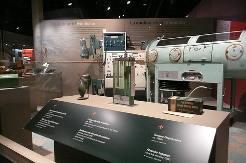
Finally, Bart Fried puts icing on the cake by adding that commercial guillotines are still sold (see for example this one from Daigger’s website) and that they can be found “in virtually every pharmaceutical company’s laboratory and in many hospital laboratories”.

Most interestingly, Bart also points to the existence of formal sets of rules and regulations for the use of such items, like the Policy and Procedures for Maintenance of Guillotines document from the University of Arizona. Well, when you think of it, of course! Foucault would smile in his grave — the governance of rat and hamster euthanasia!