Monthly Archives

May 2009

Dissection as a rite-of-passage 100 years ago — what do medical students do now?

By Biomedicine in museums

We are usually covering contemporary biomedicine on display on this blog, but sometimes older stuff gets its way into this column as well.

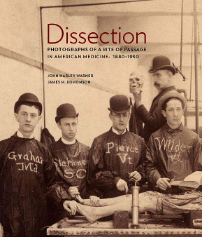
The occasion today is to draw attention to John Harley Warner and Jim Edmondson’s wonderfully illustrated book Dissection: Photographs of a Rite of Passage in America, 1880-1930, which has received very good reviews and other media attention, and has soared to Amazon rank #162.

Dissection explores a hidden treasure of images of late 19th and early 20th century medical students posing around anatomical dissection tables. The highly stylised arrangements of students, dissection tables, corpses, instruments and body parts suggest that these images were representations of a widely spread medical rite-of-passage.

To make their point, and not to be overwhelmed by too much material, John and Jim have wisely restricted themselves to medical schools in the United States around the turn of the last century — but images of similar practices probably abound in European archives and image collections as well. If I remember rightly, we have some of these in our image collection too.

There is a good interview with Jim on www.scienceFriday.com — listen to the podcast here.

Just a final thought. Now that human anatomy and dissection is no longer a core activity in medical schools — many schools have even dropped dissection on physical bodies and restrict their anatomical training to the virtual space — what kind of rite-of-passage, if any, has taken over? After all, micropipetting (a word that my speech recognition software does not recognise 🙂 is not a particularly evocative rite-of-passage practice.

Good old history of science is big news for BBC

By Biomedicine in museums

Jon Agar at UCL’s Department of Science and Technology Studies quotes an ‘intriguing announcement’ for the new series of BBC Radio 4’s Leading Edge broadcast:

For the past decade this programme’s principal concern has been with the products of science with its findings whether a freshly disinterred fossil, or a distant galaxy, a recent observation or a new theory. Starting this week we are shifting the focus from the findings themselves to the process by which they are found. Instead of treating science as an accumulating mountain of facts we will look at the who, the why, the what and the how. Who does the work? Why is it done? And how scientists operate as people who are more than the sum of their scientific publications [my emphasis]

‘Well that sounds like STS to me’, comments Jon. Well to me it sounds like good old history of science 🙂 And better late than never!

Universities and their museums

By Biomedicine in museums

The program for the Universeum Network Meeting in Toulouse, 11-13 June, has eventually been put online. Unfortunately, none of us here at Medical Museion can participate because we are opening our next big exhibition, Split and Splice, on 11 June, so we will miss contribitions like ‘Towards a university research museum?’ (Daniel Raichvarg & Marie-Laure Baudement), ‘Web archiving and university heritage: Past and future preservation, documentation and annotation of ephemeral collections for research’ (Charles van den Heuvel), ‘When the University creates intangible heritage in the 21st century’ (Marie Depraetere & Nathalie Nyst), ‘Managing the scientific and cultural heritage of a medieval university: The case of Uppsala University’ (John Worley), ‘Progressions towards establishing a Museum of the History of Science, Technology and Medicine at the University of Leeds’ (Mark Steadman), ‘The “Academic Museum” at the University of Goettingen: The University Collection as a Space of Knowledge Production and Cultural Heritage (Marian Füssel, Dominik Collet & Marie Luisa Allemeyer) — and several others. Read the full program here, and directions for attendence here.

The laboratory as an exhibition venue

By Biomedicine in museums

My friend Michael (who is a regular reader of the German HSozuKult-list) has drawn my attention to the meeting ‘Wissenschaft im Museum: Ausstellung im Labor’, to be held in Tübingen, Germany, 8-9 April 2010.

In contrast to the usual discourse about displays of science in museums, this English-German bilingual ‘Tagung’ will concentrate on the relationship between scientific practices and presentation practices in the laboratory:

Our assumption is, that this two-way relation is not only part of scientific representation, but also shows epistemological processes. Exhibitions and showrooms in scientific work spaces are not only displays of knowledge, but play a crucial role in its production. Thus, the leading question is: How much exhibition is there in science?

Interesting point! In the longer background text for the meeting, the organisers —Margarete Vöhringer at Zentrum für Literatur- und Kulturforschung in Berlin (voehringer@zfl-berlin.org) and Anke Te Heesen, Ludwig-Uhland-Institut für Empirische Kulturwissenschaft in Tübingen (anke.te-heesen@uni-tuebingen.de) — give two historical examples from the turn of the last century (Bechterev and Haeckel). But I wonder to what extent such exhibitions and displays really play a role in contemporary laboratory practice, e.g., in biomedical and biotech lab settings?

I guess the answer depends on how far the domain of the ‘laboratory’ is stretched. If, as Margarete and Anke suggest, the scientific poster session has evolved from such earlier installations in the lab, you may say that congress posters are extensions of the lab. And maybe lab visualisations online, like the application of lab protocols in JoVE, could be understood as displays in such an extended laboratory rather than communications of moving images from the lab?

Anyway, this sounds like an interesting meeting, which could bring some historical perspective on the relation between museum displays and scientific practices today, and vice versa. Send your paper proposals to Margarete Vöhringer (voehringer@zfl-berlin.org) or Anke Te Heesen (anke.te-heesen@uni-tuebingen.de) before 15 June. You can read more here.

Genomic art is so much last year

By Biomedicine in museums

Last night, I had a couple of beers with an American bioartist and some of his friends. I have always been somewhat sceptical about what is going on in so called genomic art, and after my first pint of Herslev Pale Ale, I suddenly found myself saying: ‘Genomic art is so much last year’.

My guest protested vigorously, probably because some of his own recent work can be designated ‘genomic art’. How could I mean that? Before he had the chance to refer to the many great works that have been produced in the last 10 years, I continued my argument.

Genomic art grew up in the wake of the immense public interest in the human genome project in the 1990s and early 2000s. It was an oblique response to the importance attached to the genome among scientists, funding agencies, pharma companies, the media, and social critics. Genomic art was fuelled in a tension between the bioscience establishment and a critical political and intellectual movement.

Now, the heat of genomics is over. What is now discussed in the research world — in private/public laboratories and in the board rooms of funding agencies, Big Pharma, and biotech companies — is proteomics. Largely because the design of specific, targeted protein molecules and knowledge about their interactions in the cell is supposed to have great potentials for future drug production.

Genomic art will continue to be produced, of course, but it is no longer fuelled by the political tension of the 1990s. It now lives a life of its own, less driven by political and critical discussions. However, there is so far not much interesting protein art to speak of.

My bioartist friend didn’t agree. Maybe I was right with respect to the changes taking place in the research and industrial world, but he thought I attached too much significance to this change, because proteins are the product of the transcription and translation of DNA/RNA and therefore play a secondary role on life — and, as a consequence, for critical bioartists.

To which I responded that in spite of this ‘secondary role’ of proteins, artists interested in materiality (which most bioartists are) should nevertheless pay more attention to them than to DNA/RNA, because the material substrate of the cell and body functions lies precisely in the interaction between proteins, not in the information that has coded for them.

Again disagreement: Nucleic acids are not just code, they are material too. Right you are, I said, but the material presence and direct material consequences of nucleic acids in the body is small compared to the material presence and consequences of proteins, so we can as well forget about it.

Unfortunately, our discussion ended there, because I had to go home. If I could have continued it, however, I would have said that I’m of course not suggesting that the future direction of bioart should be determined by shifting research focuses in the biosciences, or that the strong material presence of this or that molecular species should be a reason for an artist to pay attention to it. That is not how bioart, or any other art form, works.

But the fact that my bioartist friend and I got into such a heated discussion about genomic versus proteomic art is nevertheless interesting, I think.

(Dictated through my Dragon speech recognition software, therefore, alas, no links or images today)
Pas på med kemi …

By Biomedicine in museums

Det har været en rimelig fælles opfattelse herinde på Medicinsk Museion igennem en årrække at vi ikke skal være en didaktisk museumsinstitution. Opgaven er ikke primært at undervise i kroppen og dens funktioner. Vi er ikke en tredimensionel medicinsk lærebog, men et museum som skal give et historisk, kulturelt og eksistentielt perspektiv på kroppen i sundhed og sygdom. 

Men nogle gange kan man virkelig få lyst til at vende tilbage til det klassiske pædagogiske museumsideal. Som her til morgen, hvor jeg læste en artikel i Weekendavisen-Ideer af  professor, dr.med. Philippe Grandjean om hvordan vi invaderes af kemiske stoffer udefra:  ‘Vi er omgivet af kemiske stoffer’, skriver han: ‘Nogle af dem trænger ind i kroppen, fordi vi kommer til at indånde dem, spise, drikke eller røre ved dem’ (15. maj 2009, s. 12).

Grandjean ved selvfølgelig bedre når han står i sit miljømedicinske laboratorium på Syddansk Universitet. Han ved jo ganske godt, at alt vi indånder, spiser, drikker og rører ved er kemiske stoffer. Mad er ikke andet end proteiner, fedtstoffer, kulhydrater med mere, det samme gælder luften vi indånder — ilt, kuldioxid kvælstof — lutter kemiske stoffer.

Det Grandjean egentlig mener — og advarer imod — er selvfølgelig den lille mængde af artificielt fremstillede, kemiske stoffer der findes i miljøet som en uønsket sideeffekt af industriel produktion, industrialiseret landbrug etc.. De resterende 99.999 % af den mængde kemiske stoffer vi indtager til daglig er vi jo afhængige af — ellers ville vi snart dø af kvælning og sult.

Men når han skriver i avisen, falder Grandjean altså tilbage til den nemme men forkerte brug af begrebet ‘kemi’. Ved at forkorte ‘uønskede, artificielt fremstillede, kemiske stoffer’ til det mere mundrette ‘kemiske stoffer’ bidrager han til et udbredt misbrug af ordet ‘kemi’ i medierne, hvor man tit skelner mellem ‘kemi’ som noget negativt og ‘naturligt/kropsligt’  som noget positivt.

Tænk fx på alle de kosmetikfirmaer som annoncerer med at deres produkter angiveligt ikke indholder ‘kemi’ — det mener de forhåbentlig ikke, for så solgte de det rene vacuum.

Det kan synes som en rimeligt uskyldig sprogbrug. Men den slappe brug af ordet ‘kemi’ i medierne og på webben bidrager til den udbredte mangel på viden i befolkningen om, hvordan kroppen egentlig fungerer.

Organer, væv, endda celler, accepterer de fleste. Men at vi skulle være komplicerede biokemiske maskiner, hvor tusindevis af kemiske stoffer hvert brøkdel af et sekund indgår i milliarder af kemiske reaktioner med hinanden — det er ligesom ikke trængt helt igennem endnu. Og Grandjean’s kemiske invasionsmetafor gør det ikke nemmere at forklare det.

Så derfor længes jeg næsten efter det didaktiske medicinske museum igen. Med i hvert fald én lille udstilling som anskueliggør, hvordan mennesket både i sundhed og sygdom er et fin-tunet netværk af komplicerede kemiske processer. Oplysningens tid er ikke helt forbi endnu.

Sartoblot II-S — the whereabouts

By Biomedicine in museums

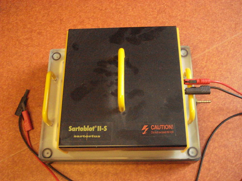
As I told in an earlier post, we are working on an exhibition about the history of proteins, which will open at the faculty of Health Sciences in early September. I visited the medical history museum in Uppsala, Sweden, a few months ago to see their astounding collections of clinical chemical artefacts. Here I found, among other things, an electrophoresis apparatus  made by the laboratory device company Sartorius — a so called Sartoblot II-S — a wonderfully coloured box which seems to have been standard equipment in biomedical laboratories in the 1980’s and 1990’s: you can still buy used specimens on the web from second hand dealers.  

The problem is that I don’t know how, when and where this kind of apparatus was used in daily practice. Does anybody have any images of it in a laboratory setting? Any gels produced by it? Any experimental results coming out of it? All kinds of info are much appreciated.

Fewer postings for a while — tendonitis, it's pretty painful

By Biomedicine in museums

There has been rather scarce posting on this site for the last couple of weeks. Several of us are extremely busy preparing the new exhibition, Split and Splice, which will open on Friday, 11 June, in our main building — hopefully, we will be able to come back with a few appetisers in the next few weeks.

But posting has also been slow because I have developed tendonitis (“mouse arm”) by using the computer mouse too much. My GP has ordered me to abstain from any sort of writing for the next month (80% reduction in working capacity). However, one on my friends in Gothenburg (Jan Nolin), who has had the problem for several years now, recommended a speech recognition software, Dragon, which I just bought and installed, and which I’m now training.

It is quite fabulous. I can write in Word, I can use Outlook — and, as you can see, I can also dictate text here into the WordPress blog (it even recognises when I say ‘WordPress’, although it cannot put the name in inverted commas, and it cannot end the sentence with a smiley, which I have to do by hand, as in the old days :-).

It has taken me three minutes to dictate the paragraph above (should be paragraphs, not paragraph, you need to be distinct when you speak to it! — and it cannot end a parenthesis for some reason). I’m trying to figure out if it can also link as well. It probably can, let’s see! The more tricky exercise will be to add pictures from Flickr (wow!, it can spell Flickr!), and to place them where I want to have them.

Does anybody have any experiences from working with Dragon in WordPress?

How to depict life itself?

By Biomedicine in museums

Just to let you know, on 12 May art historian Robert Zwijnenberg is giving a talk at the Berlin-Brandenburg Academy of Sciences and Humanities in Berlin about “How to Depict Life. A Short History of the Imagination of Human Interiority”. Here is the exciting abstract:

From 14th-century pictorial efforts to the images produced by visualization technologies, such as fMRT, the depiction of human interiority has always also been a struggle to depict and understand life itself. But how to depict interiority in such a way that life itself becomes understandable? This question was as much a problem for the anatomist of early modern times as it is for the 21st-century molecular bioscientist.

The talk will take place at 7.30 pm in the Akademiegebäude am Gendarmenmarkt, Leibniz-Saal Markgrafenstraße 38, Berlin.