
I wonder how much we could do out of photo- and electronmicroscopic art in an exhibition context? The practice of turning microscopic scientifc objects into art objects goes all the way back to the beginning of light microscopy in seventeenth century, and since then generations of microscope users have alternated between taking a scientific and an aesthetic approach to what they saw through the ocular.
So there is an enormous amount of potentially useful historical material out there. Today most scientists are accustomed to such science/art objects; almost every issue of Nature, Science, Cell, and many other scientific journals carries photo- or electronmicrographs of cellular and molecular structures on the cover.
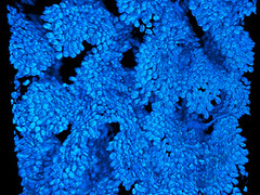
General audiences are also getting access to more and more of this kind of borderline science/art micro-iconography through popular science magazines and not least through the web. One of the best sites is Nikon’s Small World site which contains many hundreds of photomicrographs from the annual Small World Photomicrography Competition. For example, this “Cell nuclei of the mouse colon” by Paul Appleton at the University of Dundee (740x with 2-photon fluorescence microscopy; see it in context here) was the winner of the 2006 competition:
What place may such photo and/or electronmicrographs have in displays of recent biomedicine? Has anyone found a good example of an exhibition where they fit in with/complement material objects and/or texts? How can we use them to problematise the meaning-presence tension? Any views on this?