
Cell image and video library gets NIH stimulus grant

By Biomedicine in museums

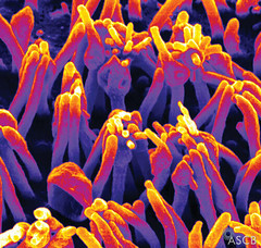
As some of you may have noticed, the online Image & Video Library of The American Society for Cell Biology has been closed since February, and nobody knew whether it would be opened again.

Last Thursday the ACSB announced, however, that the site will be re-opened and developed further by means of a $2,5 million ‘stimulus grant’ from the NIH (one of the consequences of the new Obama administration).

According to ACSB’s press release, the present image and video collection will be turned into “a comprehensive, international digital library” and furthermore, by “developing a systematic protocol for acquiring, reviewing, annotating, and uploading the images”, the ASCB will create “an efficient platform for building the library at a rapid rate”.

These are exciting news for all cell image fans!

On the boundary of visual and performative arts and biomedicine

By Biomedicine in museums

Ever noticed that the uniform resource locator (a.k.a. url) of this blog is www.corporeality.net/museion? I’ve just realised there is a url-alike in the same business as ours, namely www.CORPOrealities.org.

CORPOrealities is the website of a research project “situated on the very boundary of visual and performative arts and biomedicine”, which free-lance Viennese sociologist and artist Christina Lammer has carried out together with a team of visual artists, curators, historians and caregivers at the Medizinische Universität Wien (MUW) during the last five years.

The project is interesting in a ‘Biomedicine-on-display’-perspective because Lammer and her co-workers have used video as an ethnographic method for translating human experiences of illness and suffering into aesthetic expression. She claims that these visual ethnographic and body art interventions can “enhance complex processes of translation and mediation and strengthen the empathy, sensitivity and emotional competence in health care work” (quoted from C. Lammer, ‘Translating experience: The creation of videos of physicians and patients in the environment of an Austrian university hospital’, Int. J. of Multiple Research Approaches, vol 3: 264-75, 2009, abstract here).

Would like to see more of that kind of studies, because its feeds into the ‘Health Promotion and Innovation’ research program that we are developing together with the ethnologists here at University of Copenhagen within the frame of the Center for Healthy Ageing (see announcements for phd and postdoc positions here).

The CORPOrealities project is scheduled to end this October. Then Lammer is planning two other projects: one called ‘Features: Vienna Face Project’ and another called ‘Surgical Wrappings’. Keep an eye open!

Vi søger en publikumsmedarbejder …

By Biomedicine in museums

Medicinsk Museion søger en publikumsmedarbejder med tiltrædelse snarest muligt (HK-stilling).

Vi er et lille men dynamisk universitetsmuseum med i alt syv faste medarbejdere, der arbejder tæt sammen. Dine hovedopgaver vil være at sælge og organisere omvisninger og andre faglige arrangementer samt stå for en række administrative opgaver, herunder:

  • bookning, fakturering og kasseansvar
  • telefon- og personkontakt med kunderne/publikum
  • koordinator for museets ca. 20 studenteromvisere
  • kontor- og serviceopgaver som støtte til husets funktioner
  • medvirke til udførelse af publikumsarrangementer
  • salgsopgaver, herunder webannoncering m.m.

Du vil indgå i et tæt samarbejde med de øvrige medarbejdere, især AC-formidlingsmedarbejderen.’

Vi leder efter en person som har følgende kvalifikationer:

  • en relevant uddannelse
  • en åben personlighed og gode samarbejdsevne
  • serviceorientering, selvstændig og med ordenssans
  • behersker både dansk og engelsk i skrift og tale
  • god til Word og Excel samt lyst og evne til at arbejde med både fakturering/kasse og email-baseret
  • publikumskommunikation
  • lyst til at videreudvikle kontaktfladen med sundhedsvæsnet og det almene publikum
  • evne til at holde mange bolde i luften samtidigt

Løn og ansættelsesvilkår
I henhold til gældende overenskomst mellem Finansministeriet og Statsansattes Kartel samt Organisationsaftale for Kontorfunktionærer i Statens Tjeneste. Stillingen aflønnes i løngruppe 3, sats 1, hvor basismånedslønnen p.t. udgør kr. 21.203,17 md. ved en ugentlig arbejdstid på 32 timer.

Stillingen er en deltidsansættelse, pt. med en ugentlig arbejdstid på 32 timer. Arbejdstiden kan gøres fleksibel. Der kan også være perioder med overarbejde, som honoreres efter gældende takster.

For nærmere oplysninger, ring formidlingsmedarbejder Bente Vinge Pedersen, 3532 3821 eller museumschef Thomas Söderqvist, 2875 3801.

Ansøgning
Send din ansøgning bilagt CV samt dokumentation for uddannelse elektronisk — se link her. Vi skal have modtaget din ansøgning senest onsdag den 21. oktober 2009 kl. 12.00. Vi modtager kun elektroniske ansøgninger.

Instituttet ønsker en medarbejdersammensætning, der afspejler det omgivende samfund, og opfordrer alle interesserede uanset alder, køn, race, religion eller etnisk tilhørsforhold til at søge.

Medicinsk Museion, www.museion.ku.dk, er en del af Institut for Folkesundhedsvidenskab, Det Sundhedsvidenskabelige Fakultet, Københavns Universitet. Museet driver forskning, kulturarvsarbejde og udstillingsvirksomhed med fokus på den moderne biomedicin i samspil med kultur og samfund og rummer en af Europas rigeste medicinhistoriske samlinger i det gamle Kgl. Kirurgiske Akademi og tilstødende bygninger i Bredgade.

Waiting for the 2009 Celldance winners

By Biomedicine in museums

The art of animation of cellular and molecular processes has developed immensely in the last decade. One of the interesting trends is the increasingly sophisticated practice of mixing scientific footage with animation procedures.

A nice example is ‘The Golgi apparatus’ movie (Sougrat R. The Golgi apparatus. ASCB Image & Video Library. 2008;VID-142) that was awarded 1st Place Public Outreach Video at Celldance 2008, the annual cell film and image contest for members of the American Society for Cell Biology (ASCB), organised “to open the eyes of the world to the best in visually stunning videos and images that illuminate cell biology”. See it here: http://cellimages.ascb.org/ 

The Golgi movie animation takes you inside a mammalian cell where you can see the nucleus and its envelope that is connected to the endoplasmic reticulum (ER). Transition electron microscopy and tomography was used to create an animated image of a portion of a Golgi ribbon, where the trans-Golgi network peels off from the cisternae while a new component from the ER enters the cis-element of the Golgi. Very dynamic — very instructive — even looks nice!

This video is the first project by the Biovisualization program at the National Institute for Child Health and Human Development. Computer science and animation students collaborated with researchers to produce this visualization. As they say: “While the main focus was scientific accuracy, aesthetics were also considered”:

To convey the sense of scale, a progression is made, from actual confocal microscopy into an SEM style animation and then into non-photorealistic rendering of what can be seen at TEM level magnification. The software tools utilized for this project include Autodesk Maya, Adobe Photoshop and After Effects, Final Cut Studio, Mercury Amira and Imod. 

The winner of the 2009 Celldance contest will be announced at the ASCB meeting in San Diego, 5-9 December.

Assembling bodies

By Biomedicine in museums

Don’t forget to see ‘Assembling Bodies: Art, Science & Imagination’, the major interdisciplinary exhibition at the University of Cambridge’s Museum of Archaeology and Anthropology, when you are in town (45 mins from Stansted airport).

The exhibition explores some of the different ways that bodies are imagined, understood and transformed in the arts, social and bio-medical sciences. The project is part of the Leverhulme Research Project ‘Changing Beliefs of the Human Body’ (2004-2009), which has brought together researchers in archaeology, ancient history and social anthropology.

A series of special events and activities for a wide range of audiences is running throughout the period of the exhibition, until December 2010. For further details, see the exhibition website: http://maa.cam.ac.uk/assemblingbodies/exhibition.

Protein sculptures

By Biomedicine in museums

In the last ten years or so, in the wake of the renewed interest in protein research and proteomics after genomics, we have seen more and more artists making protein sculptures. See, for example, Graphic Thought Facility’s neon protein artwork, or Colin Rennies glass sculpture of ATP synthase, or Julian Voss-Andreae’s wood and steel sculpures of proteins, just to mention a few.

Here’s another recent example. Herwig Turk sent me these images from his current exhibition gaps (with Paulo Pereira and Johannes Hoffmann) at the Museu da Ciência, Coimbra, Portugal (the museum of the Universidade de Coimbra):

Made by ropes and epoxy and coloured with red ship paint, gaps is based on a 3D-model of connexin43 drawn by PhD-student Steve Catarino at Universidade de Coimbra in connection with a research project supervised by Paulo Pereira.

Cx43 is one of the several specific proteins that form so called ‘gap-junctions’, i.e., channels that allow signal molecules to pass the membranes between cells. Intercellular communication by means of ‘gap-junctions’ is vitally important for many bodily functions; best known is the gap junction signalling responsible for the coordination of heart beat.

Herwig explains the background for the artwork:  [Steve] did a sketch to explain it to the students and we build a sculpture after that sketch”:

Digestive history

By Biomedicine in museums

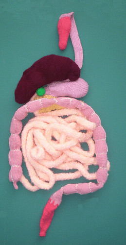
My stomach rumbled with excitement when I read the call for papers for a workshop titled ‘History, Digestion and Society: New Perspectives’ at University College Dublin, 30 April – 1 May 2010, organised at the Centre for the History of Medicine in Ireland.

As the organisers (Ian Miller and Mike Liffey) point out, diet and digestion are neglected in histories of the body, health and medicine. And diseases of the digestive system, like dyspepsia, peptic ulcer disease, vomiting etc., are not properly historically contextualised:

(image of knitted stomach from Strange but Trewe)

Meanwhile, historical analysis of issues related to food and eating often reveals a tendency to stress the political elements of historical events at the expense of the biological and medical. Topics such as hunger strikes, and the rise of organised movements such as the Temperance movement and organised vegetarianism have complex medical and biological aspects which are worthy of serious analytical attention.

Possible topics include:

  • Refusal to eat food (e.g. hunger strikes)
  • Dietary movements (e.g. temperance societies, vegetarianism)
  • The development of related technologies such as frozen food and processed food.
  • Historical concepts related to understandings of nutrition
  • The history of individual digestive organs such as the stomach
  • Medical issues related to digestion (e.g. gastric ulcer disease, indigestion)
  • Socio-cultural issues related to obesity and anorexia.
  • Surgical and medical intervention in the digestive system.
  • Human and animal digestive habits
  • Digestion and criminal activity (e.g. poisoning)

Send a 250 word abstract to Ian Miller (ian.miller2@ucd.ie) no later than 30 November 2009. For further info: Mike Liffey (michael.liffey@ucd.ie)

The colours of biomedical lab equipment

By Biomedicine in museums

If the colour of medicine is green — what is (are) the colour(s) of the biomedical laboratory? And how have these colours shifted over time?

I’m asking, because David just sent this image of a “gorgeous MacBeth densitometer” (cat.nr. 1998.0174) telling me he’s now looking around for “space age blue” in his museum’s collection.

I guess the biomedical laboratory is a more rainbowish affair. Consider this awesome Sartoblot:

(see earlier post here)

We're apparently lagging behind on the social web media side

By Biomedicine in museums

Rose Sherman (Director of Enterprise Technology at the Minnesota Historical Society) is circulating a survey about how museums are engaging their communities through social media technologies — blogs, Twitter, Facebook, MySpace, LinkedIn, Flickr, YouTube, Wikipedia, etc.

Rose asks a lot of relevant questions, like:

  • How frequently, on average, are your social media web sites updated?
  • What function(s) in your organization has staff assigned to use social media tools to engage communities?
  • How many full time equivalent (FTE) positions are assigned to use social media tools to engage communities?
  • What function in your organization is primarily responsible for actively using social media tools to engage communities?
  • On average, how many hours per week do staff spend on actively using social media tools to engage communities, e.g. they Tweet, update Facebook pages, post photos to Flickr, post videos to YouTube, etc. ?
  • Do you have a full time position(s) assigned to engaging with your social media networks?
  • Do you have a social media policy? (e.g. http://sites.google.com/site/wharman/social-media-strategy-handbook?

Thought-provoking questions which remind me that the function of such surveys is sometimes to make you aware of what you haven’t done yet. A small museum like ours is particularly provoked by questions like “How many full time equivalent (FTE) positions are assigned to use social media tools to engage communities?”.  I would rather have preferred: “How many infinitesimal part time equivalents …”.

So we’ve got lots to do on the social web media side.

You can fill in the survey here.

Sk-interfaces in extended continuation — now in Luxembourg

By Biomedicine in museums

Later today, the art exhibition SK-INTERFACES — originally displayed in Liverpool in 2008 (see earlier post here) — opens in “extended continuation” form (what others would call perpetual beta 🙂 at Casino Luxembourg in Luxembourg.

The opening event features Kira O’Reilly (inthewrongplaceness), Yann Marussich (Bleu Remix), Paul Vanouse (Relative Velocity Inscription Device) and Jun Takita (Light, only light!). The show, which is curated by Jens Hauser, is running until January 10, 2010.

Contributing artists include: Art Orienté objet, Maurice Benayoun, Zane Berzina, Critical Art Ensemble, Wim Delvoye, Olivier Goulet, Eduardo Kac, Antal Lakner, Yann Marussich, Kira O’Reilly, Zbigniew Oksiuta, ORLAN, Philippe Rahm, Julia Reodica, Stelarc, Jun Takita, The Office of Experiments, The Tissue Culture and Art Project, Sissel Tolaas, and Paul Vanouse.

Here’s the perpetual beta flyer:
Skin is our natural “interface” with the world – more and more, however, technological extensions are taking over its role; “interfaces” create both new freedoms and new constraints. In the cross-disciplinary exhibition sk-interfaces, twenty international artists reflect on how modern technosciences have altered our relationship with the world: telepresence, digital technology, speculative architectures, bio-prostheses, tissue culture or transgenics – for the artists, they are not mere topics but tools, methods and media to appropriate. They test the permeability of the borders between disciplines, art and science. Their interfaces connect us with other species, put satellite bodies up for debate, destabilize our conception of what it means to be human today, and create evolutionary scenarios confronting the technological pressure to adapt and its socio-political implications. As a natural inventor of the artificial, Homo Sapiens compensates for its imperfections through the use of technology. Arguing for the naturalness of the media created to this end, theorist Marshall McLuhan once suggested that they be understood as bodily extensions per se – something not unlike an electronic skin spanning the world in which inner and outer were no longer clearly distinguishable. Yet, these prosthetic extensions come at the high price of “auto-amputation”, for each prosthesis permits other senses and states of consciousness to be numbed and to atrophy. Today, in the context of the so-called Life Sciences, media and technological interfaces can no longer be considered merely as telecommunicative, digital, or human-machine interfaces; in the age of bio-facticity, even that which apparently grows naturally is now technologically induced, producing biological artefacts. In view of the utopias and dystopias this inspires, it is no surprise that artists take up the material, function and metaphor of skin as the original, semipermeable and active membrane. They contest the predominating utilitarianism with subversive alienation, aesthetically, poetically and provocatively. Sometimes they wrest from the technological a holistic impulse, sometimes an ecological illusion in which humans admit their responsibility rather than isolate themselves in their alleged superior status. Hence, sk-interfaces examines above all the “ – ”: the in-between-space of our contemporary ontological grey zones.