
The good old History of Science Society (HSS) and most other history of science, technology and medicine meetings continue the tradition of organising endless rows of parallell sessions, each with the standard 3-4 papers á 20 minutes + concluding Q&A. But some societies are trying something new. Just got the call for next year’s Swedish history of science and technology meeting (Teknik- och vetenskapshistoriska dagar) in Gothenburg in November 2010. The conference is going to be held in the regional science park (Lindholmen Science Park), the organisers are downplaying traditional papers sessions and are instead intent on creating a more participatory-driven meeting, with a greater variety of interaction formats. Unfortunately in Swedish only, I’m afraid, but that aside it seems like the Swedes are keen on breaking up the traditional conference format. More info from Lena Ewertsson at the Dept of Technology and Science Studies, University of Gothenburg, lena.ewertsson@sts.gu.se. See also http://www.sntv.kva.se/files/2010_1st_CALL_FOR_PAPERS.pdf.
In late March, Rikke Schmidt Kjærgaard (which several of us here at Medical Museion met when she gave a seminar here a couple of years ago and who is now working at the MRC Mitochondrial Biology Unit, University of Cambridge) is organising a meeting of great relevance for anyone interested in biomedicine on display, whether in museums or on the screen.
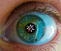 Titled ‘Have you ever seen a molecule? Art, science and visual communication’, the two-day meeting at the Cambridge Centre for Research in the Arts, Social Sciences and Humanities (CRASSH), 25-26 March, concentrates on the correlation between art/design and molecular biology, in particular structural biology, and on the impact of the arts and artistic practices on scientific culture. Current molecular biological research is very dependent upon visualisation methods, both in the production of intepreted data and in the communication to other scientists and the public at large. The call for papers explains the relevance of this topical issue, both for scientists and for science communicators, understood broadly:
Despite the fact that structural images of individual projects are made by thousands of researchers in laboratories around the world, there is as yet no general consensus on what makes a good image. Consequently, there is no obvious and necessary correlation between the images made for pragmatic and heuristic purposes in the laboratory, those chosen for posters and conference presentations, the images accompanying article submissions, and finally those that will be selected or further designed for public engagement and communication. Instead, how specific traits should be visualised, which colour schemes should be applied and how to pick the perfect image for specific purposes depend to a large degree upon pragmatic categories and local factors within individual laboratories and research groups, as well as on editorial decisions and a stronger promotional value, at least to some degree independently of scientific preferences and arguments.
Interdisciplinary collaboration in visualising molecular structures lies at the very core of contemporary research processes and products. Bringing art, design and science together is far more than just an interesting experiment in transdisciplinary cross-communication, it is a necessary step in exploring new ways of optimising imagery at the molecular level and thus breaking new ground. We depend upon this in the arts as well as in the sciences in the future university to make things better and to advance our knowledge of life at a molecular level.
Rikke/CRASSH welcomes submissions for presentations broadly within visualisation of science. Send a <250 words abstract, a brief CV and a few lines about your interest in the conference before 1 February 2010 to rsk@mrc-mbu.cam.ac.uk (and please use the form here).
Registration fee (includes catering) is a bargain (£30 for faculty, £15 for students.). Registration will be available from the conference website shortly.
Today (Nobel Day!), Thursday 10 December at 8pm, Obervatory/Morbid Anatomy in New York hosts a talk by Mütter Museum‘s new director Robert Hicks, titled “Exquisite Corpses: Illustrated Lecture & Artifacts from the Mütter Museum”. I guess it’s too late now to get on the morning flight (unless you borrow Air Force One which stands idle on the ground in Oslo today), but the abstract might be interesting to read anyway — not least for museums that are planning to rearrange their anatomical collection (as we are):
Images of post mortem human remains are fascinating and disquieting. They amuse children at Halloween and disturb adults when on display at museums. Today’s omnipresent imagery of people doing everything at all times has not accustomed us to depictions of human mortality. The dead are speedily removed from view, and our direct contact with the dead is limited and controlled. Although mortal images can arouse empathy and may develop tolerance for a spectrum of human physical variation, other cultural voices argue for proscription and censure. In this presentation, Robert Hicks, director of the Mütter Museum explores our dialogue with post mortem human imagery by examining its relationship to politics and ownership of the dead. He incorporates perspectives drawn from anthropology, art criticism, history, museum curatorship, and criminal justice.
European Association of Museums for the History of Medical Sciences (EAMHMS) afholder sin 15. konference i København den 16. – 19. september 2010 under temaet “Contemporary medical science and technology as a challenge for museums”.
Konferencens fokus er den udfordring, den nutidige udvikling inden for biomedicinsk forskning og medicinsk teknologi stiller museumsverden over for.
Citat fra konferenceannoncen:
The image of medicine that emerges from most museum galleries and exhibitions is still dominated by pre-modern and modern understandings of an anatomical and physiological body, and by the diagnostic and therapeutical methods and instruments used to intervene with the body at the ‘molar’ and tangible level – limbs, organs, tissues, etc.
The rapid transition in the medical and health sciences and technologies over the last 50 years – towards a molecular understanding of human body in health and disease and the rise of a host of molecular and digital technologies for investigating and intervening with the body – is still largely absent in museum collections and exhibitions.
As a consequence, the public can rarely rely on museums to get an understanding of the development and impact of the medical and health sciences in the last 50 years.
Læs mere på http://tinyurl.com/ylx5atx eller kontakt Thomas Söderqvist, ths@sund.ku.dk.
The 15th biannual conference of the European Association of Museums for the History of Medical Sciences (EAMHMS) will be held at the University of Copenhagen, 16–18 September, 2010.
This year’s conference focuses on the challenge to museums posed by contemporary developments in medical science and technology.
The image of medicine that emerges from most museum galleries and exhibitions is still dominated by pre-modern and modern understandings of an anatomical and physiological body, and by the diagnostic and therapeutical methods and instruments used to intervene with the body at the ‘molar’ and tangible level — limbs, organs, tissues, etc.
The rapid transition in the medical and health sciences and technologies over the last 50 years — towards a molecular understanding of human body in health and disease and the rise of a host of molecular and digital technologies for investigating and intervening with the body — is still largely absent in museum collections and exhibitions.
As a consequence, the public can rarely rely on museums to get an understanding of the development and impact of the medical and health sciences in the last 50 years. Biochemistry and molecular biology have resulted in entirely new diagnostic methods and therapeutic regimes and a flourishing biotech industry. The elucidation of the human genome and the emergence of proteomics has opened up the possibility of personalised molecular medicine. Advances in the material sciences and information technology have given rise to a innovative and highly productive medical device industry, which is radically transforming medical practices. But few museums have so far engaged seriously and in a sustained way with these and similar phenomena in the recent history of medical sciences and technologies.
The contemporary transition in medical and health science and technology towards molecularisation, miniaturisation, mediated visualisation, digitalisation and intangibilisation is a major challenge for the museum world; not only for medical museums, but also for museums of science and technology, and indeed for all kinds of museums with an interest in the human body and the methods for intervening with it, including art museums, natural history museums and museums of cultural history.
Contemporary medicine is not only a challenge to exhibition design practices and public outreach strategies but also to acquisition methodologies, collection management and collection-based research. How do museums today handle the material and visual heritage of contemporary medical and health science and technology? How do curators wield the increasing amount and kinds of intangible scientific and digital objects? Which intellectual, conceptual, and practical questions does this challenge give rise to?
The meeting will address questions like (but not limited to):
- How can an increasingly microanatomical, molecularised, invisible and intangible (mediated) human body be represented in a museum setting? Does the post-anatomical body require new kinds of museum displays?
- How can museums make sense of contemporary molecular-based and digitalised diagnostic and thereapeutic technologies, instrumentation and investigation practices in their display practices?
- How can museums make use of their older collections together with new acquisitions from contemporary medicine and health science and technology?
- What is the role of the visual vs. the non-visual (hearing, smell, taste, touch) senses in curatorial practice and in the public displays of contemporary medical science and technology?
- What can museums learn from science centers, art-science event venues etc. with respect to the public engagement with contemporary medical science and technology? And, vice versa, what can museums provide that these institutions cannot?
- How can museums draw on bioart, ‘wet art’ and other art forms to stimulate public engagement with the changing medical and health system?
- How does physical representations of contemporary medicine in museums spaces relate to textual representations in print and digital representations on the web?
- How can museums integrate emerging social web technologies (Wikipedia, Facebook, Twitter, blogs, etc.) in the build-up of medical and health exhibitions?
- What kind of acquisition methods and policies are needed for museums to catch up with the development of contemporary medical science and technology, especially the proliferation of molecular and digital artefacts and images?
- What kind of problems do museum encounter when they expand the acquisition domain from traditional textual, visual and tangible material objects to digital artefacts (including software, audio- and videorecordings, and digitally stored data) and non-tangible scientific objects.
- How can participatory acquisitioning, crowd-sourcing, wiki-based methods, etc. (‘museum 2.0’) be employed for the preservation and curation of the contemporary medical heritage?
- How can curatorial work in museums draw on medical research and engineering and on academic scholarship in the humanities and social sciences? And, vice versa, how can museums contribute to medical teaching and research and how can their collections stimulate the use of physical objects in the humanities and social sciences?
The conference will employ a variety of session formats. In addition to keynotes and sessions with individual presentations of current research and curatorial work there will also be discussion panels and object demonstration workshops.
We welcome submissions from a wide range of scholars and specialists — including, for example, curators in medical, science and technology museums; scholars in the history, philosophy and social studies of medicine, science and technology; scholars in science and technology studies, science communication studies, museum studies, material studies and visual culture studies; biomedical scientists and clinical specialists; medical, health and pharma industry specialists with an interest in science communication; engineers and designers in the medical device industry; artists, designers and architects with an interest in museum displays, etc.
We are especially interested in presentations that involve the use of material and visual artefacts and we therefore encourage participants to bring illustrative and evocative (tangible or non-tangible) objects for demonstration.
The meeting will begin on Thursday 16 September (noon) and end on Saturday evening 19 September, 2010.
100-300 word proposals for presentations, demonstrations, discussion panels, etc. shall be sent before 28 February 2010 to the chair of the program committee, Thomas Soderqvist, ths@sund.ku.dk.
A meeting website for registration and hotel bookings will be established in early January 2010. A number of hotel rooms will be prebooked.
Programme committee:
Ken Arnold, Wellcome Collection, London
Robert Bud, Science Museum, London
Judy Chelnick, National Museum of American History, Washington, D.C.
Mieneke te Hennepe, Boerhaave Museum, Leiden
Thomas Soderqvist, Medical Museion, University of Copenhagen (chair).
Local organising committee:
Anni Harris, Bente Vinge Pedersen, Carsten Holt, Morten Bulow and Thomas Soderqvist, Medical Museion, University of Copenhagen.
For further information about the academic programme, please contact Thomas Soderqvist, ths@sund.ku.dk. For practical information about travel, accommodation, etc., see http://www.mm.ku.dk/sker/eamhms.aspx, or contact Anni Harris, konference2010@sund.ku.dk after 4 January 2010.
The conference is hosted by Medical Museion; further information will be posted on the museum’s website (www.museion.ku.dk) and on this blog.
The ICOM subcommitte on University Museums and Collections (UMAC) has set up a moderated list to facilitate exhange of information between university museums. The list is open also to non-UMAC members. See more here — for subscription, go to: https://listes.u-strasbg.fr/sympa/unistra.fr/info/umac-ml
My blood pressure rose this morning when I read Google CEO Erik Schmidt’s rebuttal of Rupert Murdoch’s attack on Google (published 1 December as an op-ed in Wall Street Journal, of all places).
Not because he strikes back at the old newspaper dinosaurs. I don’t mind, I hardly read paper media any more. The reason for my momentarily increasing pulse rate is Schmidt’s opening lines: “It’s the year 2015. The compact device in my hand delivers me the world, one news story at a time […] the device knows who I am, what I like, and what I have already read (my emphasis).
I don’t actually mind about the privacy thing. There is too much info for the new Leviathan to crunch already, so I don’t care about the surveillance problem. What really irritates me is the idea that a search machine is supposed to deliver “what I like and what I have already read”. Fine, if I opt for that possibility — but I also want to be able to opt out. Sometimes I browse to find what I know I want to find — but often I browse to find something I didn’t know I wanted to find — something that shakes me out of the expected. I want to be surprised!
It’s like falling in love: meeting someone you’ve never dreamed of. It’s like science: detecting phenomena you never expected. It’s like visual art: viewing the world in a totally unanticipated way. It’s like literature: exploring news ways of putting human experience into words.
I’m not the only one who wants to opt out; the problem has been raised several times before. But the fact that Google’s CEO unwittingly repeats the old formula for web search in his 2015 vision scares the hell out of me. Enough to put me in a state of alarm this otherwise tranquil Tuesday morning in Copenhagen, where the climate meeting has just begun only two kilometers from where we live.
One of the challenges for a museum of medicine intent on collecting recent and contemporary medical artefacts is to get an overview of the historical development of medical instruments, medical technological systems and the medical device industry.
Trade shows and their catalogues (published or online) are excellent sources. But memoirs and reminiscences of people who have been engaged in the trade show business can also be useful — they add a more personal perspective to the dry historical data, they are more fun to read than catalogues, and you can probably construct a useful picture of trends by piecing their more or less idiosyncratic stories together.
Take for example Wolfgang Albath, a pioneer in laboratory medicine and one of the founding organisers of the world`s largest medical trade show, MEDICA in Düsseldorf,. He has just summarized, shortly, his view of some of the important trends in the last 40 years of medical hospital technology (in the 12 Nov online issue of European Hospital):

Medica trade show 1974
In summary, his view of the recent history can be described in three words: mechanisation, automation and digitalisation. When MEDICA started (in Karlsruhe) in 1969, it focused exclusive on laboratory diagnostics. Most lab analysis were then carried out manually and in pretty small series.
One of the few automatic systems was the Technicon Auto-Analyzer, introduced around 1960; for a contemporary evaluation of it, see here): “Based on a system of continuous flow analysis [the Technicon AA] revolutionised lab diagnostics and paved the way for analysers to work through organ-specific parameters in batches”.
In the 1970s came immunofluorescent techniques for detecting auto-antibodies and infectious agents, and in the 1990s advances in molecular biology opened new diagnostic opportunities at the picomolar level.
Iinformation and communication technology has not only made possible automation in the clinical lab, but all kinds of hospital practices. The first patient monitoring systems, which are now taken for granted in intensive care and neonatal unit, were introduced in operating rooms and wards in the mid-1960s. In the clinical laboratory, computer development made possible large-scale diagnostic tests in the 1970s.
Another area which depends heavily on IT is radiology and medical imaging. In the 1960s “the triumph of real-time ultrasound diagnostics began”; in the 1970s came the CT-scanner; the first digital image archives, radiology information systems and laboratory information systems arrived in the mid-1980s; about the same time came MRI, and in the 1990s PET. 3D reconstructions of CT, MR and ultrasound images also became possible in the mid-1990s.
Surgery too has undergone enormous technological changes; eg., keyhole (laparoscopic) surgery began in gynaecology in 1969; the first keyhole gallbladder removal was performed in 1985 and in the early 1990s keyhole surgery in the abdomen. And then there is laser technology which has “lit up the medical sky” for 30 years, not least in ophthalmology, where doctors hardly cannot imagine work without lasers today.
While we are waiting for the sequel to Joel Howell‘s seminal Technology and the Hospital: Transforming Patient Care in the Early Twentieth Century (Johns Hopkins University Press, 1996), reminiscences like Albath’s are among the best ways to get an overview of the complexities of the recent history of medical technology. I haven’t made a systematic search for memoirs and reminiscences of similar kinds — but I’m convinced there are many out there, although they can be difficult to find.
(Btw, for a useful academic course syllabus for the history of medical technology, see here).
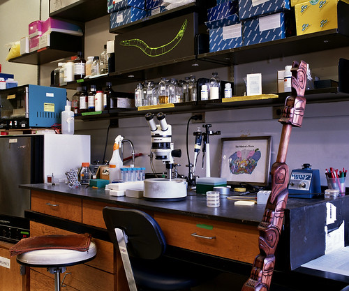
 Seed is running a series of monthly portraits of workbenches of interesting people (like Oliver Sacks, a renowned bat expert, an industrial designer, etc.)
The latest portrait, published in yesterday’s online issue, is the lab bench of Martin Chalfie, one of the three who won a medical Nobel last year for the discovery of green fluorescent protein (GFP).
The image on seedmagazine.com is interactive (of course) — that is, you can blow up details with accompanying texts.
Nifty, but …. what struck me when I first saw the image was that Chalfie’s lab bench doesn’t look authentic. Take a look at the magnified version below — it is way too neat and tidy! It looks like the photographer has cleaned up and arranged everything in orderly fashion before shooting the image.
Then I read the caption to the small glass bottles detail on the shelf above the microscope — it explains why:
I have to admit, I haven’t done a lot of experiments recently. I spend most of my time in my office next door, working on papers or talking with post-docs about their studies.
That’s the fate of most senior scientists — and Seed doesn’t seem to have realised that this fact corrupts the authenticity of the image. The difference between a used and not-so-much used lab bench is subtle. But it is there. Maybe they could have presented it as ‘the dead workbench of Martin Chalfie’ instead.
So, please, in the forthcoming issues, let’s get some images of lab workbenches that reflect some real lively untidy 24/7 lab work.
(thanks to Bertalan Meskó for the tip about Seed‘s article; that said, however, Bertalan wrongly, in my view, believes that the image “lets you look behind the scenes of the workbench of a famous and successful scientist”. That’s exactly what it does not — it’s lets you see pure surface, no behind.)
Gott exempel på forskningsformidling som tilfredsstiller alle nyhedsverdens krav på “lige her og nu!” reality show: The Brain Observatory ved University of California San Diego viser på streaming video hvordan de laver histologiske tyndsnit af hjernen af en patient. Og de gør det altså lige nu.
Patienten (som kaldes H.M) led af alvorlig amnesi. Efter hans død blev hjernen frosset ned og er — lige nu — ved at blive skåret i tynde skiver i en helorgans-mikrotom i en lang session som vil vare ca. 30 timer og som slutter en gang i aften. Hele proceduren bliver altså streamet, se her.
Så her langt var de kommet kl. 9.30 da jeg lavede en screen-dump:
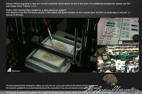
Fascinerende histology-live! Og fascinerende eksempel på forskningsformidling som lever op til “lige nu”-syndromet. The Brain Observatory gør det primært for at skaffe penge til deres forskning. Så det er led i en reklamekampagne.