Monthly Archives

March 2008

The auditory space of contemporary medicine

By Biomedicine in museums

Browsing Øystein Horgmo’s blog about medical videography (see earlier post here) I also fell over his description of the sounds of the operating room.

The auditory space is one of those forgotten dimensions of medical science studies (see an earlier post here). As far as I remember, I haven’t seen any historical work on contemporary medicine or any medical science studies project that pays attention to the medical soundscape.

That’s a shame, because the hearing sense is more open for presence-effects (á la Gumbrecht) than vision. In other words, the auditory qualities of the medical world is a potentially very interesting sensory dimension for curators of medical museums to work with.

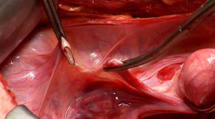
Here is Øystein’s description of the sounds of the operating room (my underline of auditory indicators; wish he had put an mp3-file on his blog instead of putting it in words!):

Below all the other sounds is the constant, but barely audible hiss of the ventilation. This is especially strong and effective in ORs, to keep the air as clean as possible. Slightly above lies the perpetual mechanic breathing of the respirator. Adding to this basis is the more prominent, but still subtle machine hum of the suction pump. It’s a sound you don’t notice till it’s gone. Nothing creates a calm atmosphere like turning off the suction. Against this constant background noise comes the busy but calm sounds of the actual surgery. The clanking of steel against steel as the scrub nurse puts instruments back on the back table. The high pitched tone from the electrocautery unit when the surgeon steps on the remote pedal. The characteristic two or three clicks of the toothed locking mechanism as a clamp is closed. The slurping sound of the suction in use. And around all this, the murmur of the surgical team. The scrub and circulating nurses exchanging equipment and counting sponges. The surgeon calling for instruments and ordering the assistant. The laughs. The small talk. This steady rhythm is broken now and then by the occasional blips of the surgeon’s pagers (left by the entrance), ringing of the intercom and the insistent beeping of the anesthetist’s monitors. And that’s about it. The aural atmosphere of the OR.

I’m already imagining up a museological research project on the representation of medical soundscapes in collections and displays. Anyone out there who would like to work with us on this topic?

Medicine on blog display: opening the black box of surgery and anasthesia

By Biomedicine in museums

Only a few years ago most bloggers were happy, omnivorous amateurs who wrote about anything that happened to pass by their computer screens. Now more and more professionals are discovering the networking powers of the medium. I’m particularly intrigued by the rapid emergence of blogs from all over the medical world—it’s like a prairie fire! Consider, for example, this list of blogs by surgeons and anaestesiologists (taken from SurgeXperiences):

Admittedly, they range from serious institutional blogs to somewhat ridiculous first-person ramblings, but this is part of the charm and strength of the genre. Together these first-hand reports give an amazing insight into the personal and social world of medicine, in this case surgery/anaesthesia. By opening up the otherwise black (or green?) box of the surgical profession, these and many other blogs constitute an important addition to the published sociological, contemporary-historical and autobiographical literature of the field, and a increasingly important source of inspiration for science studies scholars.

'The Sterile Eye': Cancer diagnostics and therapy on video display

By Biomedicine in museums

The genre of medical blogs grows incessantly, and it’s difficult to keep up with all exciting new quality start-ups. So I have to restrict mention to those that may have some medical museum relevance.

Yesterday I fell over The Sterile Eye: Life, death and surgery through a lens, edited by medical videographer Øystein Horgmo at the Norwegian National Hospital (Rikshospitalet) in Oslo, who makes clinical videos of medical and surgical procedures. For the last three years he has documented cancer diagnostics and treatment; he’s currently working on sarcomas, skin cancer and melanoma, and will continue with tumors of the liver, pancreas and other endocrine organs later this year. He has just told me that much of this material is available on the net (here)—a fine repository of clinical videos which I suppose can be used (with permission) in both physical and digital museum exhibitions (so far only in Norwegian, but an English version is underway).

Øystein Horgmo says he started The Sterile Eye in November last year because he wanted to share his experiences of videofilming diagnostic and surgical procedures to a larger public: “Being present when people are at their most vulnerable, like at the CT for detecting cancer or when in deep narcosis during surgery, is a special and strange priviliege”.

I particularly like this blog because this is not just the usual recycling of stuff from other websites, but original observations based on his professional experience as a clinical photographer and comments on the genre of medical videography.

For example, I’ve never thought of the problems involved in rendering shades of red on video. Because of the ubiquitous presence of blood throughout our body, most organs and tissues are coloured in different shades of red:


(round ligament; ligamentum rotundum; credit: ‘Different shades of red’)

When filming operation procedures for clinical instruction purposes it is therefore crucial that the video system can reproduce the colours accurately. The popular HDV video format cannot reproduce the different shades of red satisfactory, so Øystein Horgmo shoots his videos on the so called DVCPro50 format instead (technical explanation here).

A great medical blog for anyone with an interest in the history of contemporary medicine, medical science studies or medical museology. I’ll add it to my RSS feeder immediately.

Biomedical image fatigue

By Biomedicine in museums

As I wrote earlier today, the 2008 Wellcome Image Awards (formerly the Biomedical Image Awards) have been announced. 22 dazzling, advanced-tech produced, coloured images of tissues, cells and molecular models were put on display in the Wellcome Collection foyer yesterday and have also been laid out on their website for the public eye to admire.

The top of the biomedical image pops? Or what? Am I the only person who is beginning to feel saturated with biomedical images? Not only is this culture as a whole swamped with pictures—on billboards, in newspapers, on websítes and blogs, not to speak of the pictorial explosions on Youtube and Flickr. The professional biomedical media are also rapidly becoming heavily visualized. Every life science journal with self-respect puts eye-popping bio-pictures on its covers; and the articles between the covers are filled with micrographs and visualizations. The popular science media are no exception: amazing picture of dendrites, ribosomes and embryos everywhere.

Fascinating, yes! But also a trifle difficult to keep up with. The more pictures there are to see, the less time and attention each and everyone of them gets. I must admit I feel mildly saturated. Biomedical picture fatigue is beginning to set in.

Youtube, Flickr and hundreds of institutionally based online image collections are what they are, i.e., repositories. But the selection of award winners doesn’t necessarily have to result in yet another repository. So my suggestion for the next year’s Wellcome Image Award is: Be more selective! Allow the jurors to choose one picture only (maybe three at the most). And don’t take the easy solution, by leaving it to the website visitors’ vote.

My sense of fatigue also has to do with the uncritical presentation of the selected images. For sure, this is not just a problem with Wellcome Image Awards. Most awards of this kind (and there are several of them) suffer from the same problem. It is as if the jurors believe that most members of the public are still (in 2008) satisfied with yet another Photoshop-edited and colour-enhanced scanning micrograph of a flowerbed of cancer cells.

Instead of being bombarded with albums of beautiful pics, I would like to see more aesthetic assessment. Instead of just displaying their choice, the jurors should come out of the aesthetic closet and pass some outspoken critical judgement. Give us some arguments pro and contra the chosen image. What makes this select image a good picture?

My image fatigue is not alleviated by the fact that the jury has included some fairly dated pictures for the awards. For example, this image:


“Wholemount staining of an 11.5 day old mouse embryo showing parts of the nervous system stained green with an antibody to neurofilament, the floorplate and endoderm stained blue with an antibody to HNF3beta and the heart stained red. Unstained tissue appears grey” (James Sharpe, MRC Human Genetics Unit, 2003).


—produced by a technique called optical projection tomography developed at the MRC Human Genetics unit in Cambridge—was cover embryo on Science magazine already six years ago (in the April 19 issue, 2002)!

I would probably become more alert if I weren’t served off with some old and widely spread images. In fact, the well-known model of a prokaryotic ribosome which has been travelling with the University of Sunderland Design4Science exhibition (lots of good pics from the exhibition on Flickr here) is also among this year’s 22 selected award winners. I believe an image contest becomes more exciting if choice falls upon a picture we haven’t seen before!

Sk-interfaces in Liverpool a great succes — two more weeks to go

By Biomedicine in museums

Just want to remind all our readers about Jens Hauser’s skin-exhibition in Liverpool (see earlier post here) which is breaking the visitor records at FACT/Liverpool and ends with a special closing event on 29 March. Furthermore, the conference that went with the exhibition is now on record; and finally, the book (sk-interfaces: Exploding Borders–Creating Membranes in Art, Technology and Society) is out.

See much more details below (unedited material for connoisseurs): Read More

2008 Wellcome Image Awards — biomedical pictures galore

By Biomedicine in museums

This year’s Wellcome Image Awards were announced Tuesday night. The 22 images chosen by a jury from the huge collection in the Wellcome Library’s image repository (Wellcome Images) will be shown in the Wellcome Collection foyer at 183 Euston Rd., London, until some time this summer—and then at the leading Japanese science center, Miraikan (The National Museum of Emerging Science and Innovation) in Tokyo.

It’s an amazing feast for they eye; showing an array of images of tissue, cellular and molecular structures produced by a variety of impressive imaging technologies, displayed in dazzling enhanced colours—like this scanning electron micrograph of prostate cancer cells by Annie Cavanagh at the Multimedia Unit of the School of Pharmacy, University of London:

 And there are many more here.

Despite all this impressive biomedical beauty, I have some critical remarks, both about the selection of images, and the genre as such. Will be back later tonight when I have sorted out my views.

Oral history, caring, health and illness

By Biomedicine in museums

Oral history is strong in the UK, and so is history of medicine. The topic for this year’s Oral History Society‘s Annual Conference is ‘Who Cared? Oral History, Caring, Health and Illness: Marking 60 Years of the National Health Service’. The meeting is organised in association with the University of Birmingham Centre for the History of Medicine and takes place in Birmingham 4-5 July. More info here. Seems like a good opportunity to refuel the oral history skill tanks.

What is 'the inclusive museum'? Part 2

By Biomedicine in museums

I think Sniff raised some timely questions in her post last Friday about the upcoming International Conference on the Inclusive Museum (to be held in Leiden in early June)—especially what the meaning of the buzz-word ‘inclusion’ is, and why museums should be ‘inclusive’.

The ‘Scope and Concerns’ page of the conference website is an intriguing programme document. As I read it, the organisers’ argument for ‘the inclusive museum’ runs approx. as follows:

  1. ‘this time of fundamental change’ asks for new roles for museums.
  2. there is no universal visitor any more (at least not in the ‘old’ sense).
  3. on the contrary, museums visitors are a diverse crowd, and there are many dimensions of diversity.
  4. how does then participation in museums work?
  5. the answer is to be open (inclusive) to all these forms of diversity—which in turn involves two things:
  6. first, the recognise the particularity of visitors,
  7. and, second, to create new forms of universality, “where every visitor is allowed the space to create their own meanings, where no visitor is left out”.
  8. this new inclusivity involves new forms of engagement which introduces new forms of active participation on the side of ‘visitors’: “a blurring of roles, between producers and consumers of knowledge, between creators and readers of culture, and between the person in command and the person consenting”.
  9. the new inclusivity also involves new modalities of representation, especially those based on digital technologies.

What they basically say, I think, is that museums need to cater for a more diverse crowd of demanding visitors, and that this necessitates a more creative use of digital media technology.

Not a particularly revolutionary conclusion! But what’s more interesting is, to relate to Sniff’s questions, why this process is described in terms of ‘the inclusive museum’. Is this really the best way to conceptualise what is going on in ‘this time of fundamental change’ (globalisation)?

Isn’t the current change in the role of museums better understood in terms of ‘marketization’? In other words, instead of thinking of globalisation as a process of cultural ‘inclusivity’, isn’t the changing role of museums better conceptualised in terms of their economic transformation—away from being state institutions in which academic and curatorial elites control the collections and exhibitions, and towards new forms of market-oriented corporations? Rather than serving a diversity of ‘particular visitors’, aren’t they becoming more oriented to the needs of consumers, using the best available sales and advertisement methods available, including digital technologies?

I believe marketization has a lot of positive consequences for museums (dont’ forget that many museums, not least art museums, have always operated on market conditions with excellent results). There are many good reasons to open up for a more two-way communication between buyers and sellers of cultural experiences, and to open up for a larger degree of reciprocality in the production and appreciation of cultural heritages (consumer influence). And nobody would probably disagree that digital media technology is here to stay (cf. museums and web 2.0).

But there are also negative consequences of this marketization, for example, a profound risk for lowering the quality of museums, especially in cultural history museums which are not used to operate on the market. And I believe these negative consequences are better understood and opposed if one avoids the fluffy notion of ‘the inclusive museum’, and instead takes a more unsentimental and realistic view of what is going on in the museum world.

Acquisitions are the lifeblood of museums

By Biomedicine in museums

Formerly announced workshop ’Communicating Medicine: Objects and Objectives’—held Friday 7 March at the Centre for History of Science, Technology and Medicine (CHSTM) in Manchester—gathered over 40 scholars and curators, mainly from the UK.

There were nine presentations in all. One each from Science Museum (London), Museum Boerhaave (Leiden), the Wellcome Collection (London), and the Sedgwick Museum (Cambridge), and another five from us here at Medical Museion (Copenhagen): by Søren Bak-Jensen, Susanne Bauer, Jan Eric Olsén, Camilla Mordhorst and myself (see full programme here and here).

 (Susanne Bauer)

Altogether this was a varied and inspiring day about medical museum exhibitions and collections. I’m afraid I was a trifle too involved in the discussions to be able to give a fair resumé of what went on. Suffice it to say I was particularly concerned with Francis Neary’s (Sedgwick Museum) contribution, because Francis brought up the notion of ‘things-that-talk’ in connection with his (otherwise beautifully crafted) argument about machines and instruments as agents.

 (Francis Neary)

As readers of this blog may have noticed, Adam and I have recently had some serious doubts about the usefulness of the ‘things-that-talk’ metaphor (see here, here and here), so Francis’s argument gave rise to some critical questions in the discussion that followed. Why impute agency to instruments? What do we gain from doing so?

Also raising lot of discussion was Søren’s paper on collecting biomedicine and the experiences of acquiring contemporary biomedical artefacts during the University of Copenhagen Medical Faculty Garbage Day last June


(Søren Bak-Jensen)

Søren’s presentation made me think of former British Museum Director Robert Anderson’s point that ‘acquisitions are the life blood of museums’. Or to put it another way: research can be seen as the soul of museums, and exhibitions their public face and rationale for public funding—but the incessant acquisition of new artefacts provides the life-sustaining nourishment for museum institutions.

I’m not sure that all medical historians or medical museum curators today are fully aware of the consequences of Robert Anderson’s wisdom. So next time we meet we should perhaps discuss how to collect medical objects rather than how to use them for communicating medicine?

 (John Pickstone listening attentively)

Altogether a most enjoyable day, well worth the trip and air traffic delays, and very well organised by CHSTM’s outreach officer, Emm Barnes:

Btw. did anyone else take better notes than I did?