After the minisymposium with Jens Hauser and Sepp Gumbrecht on the concept of ‘presence’ here at Medical Museion last spring, our research group has repeatedly come back to the relation between mediated visualizations of biomedical objects, on the one hand, and the immediacy of touching them, on the other (see, for example, Jan Eric’s earlier post on Condillac’s statue).
As an anecdotal illustration of the immediacy of touch, I’d like to present the following personal experience.
In mid-March, I accompanied my partner to the National Hospital here in Copenhagen for an ultrasound scan of our then 13 week old foetus. As thousands of other prospective parents we were of course thrilled by what we saw on the screen:
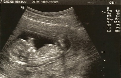
Watching our future baby ‘live’ on a computer screen like this was quite amazing, even though we had seen such pictures on the internet before. It’s an experience we share with millions of others: digitalized ultrasound scanning foetus images have become an integral part of the contemporary understanding of what it means to deliver new citizens to the world. A striking image of early life that is easily communicated in our visual culture and as such an illustration of the formation of biocitizenship, both discursively and substantially.
Back to the anecdote: Six weeks later, we went for the second screening and watched the same kind of picture, just more detailed, with ears, fingers, toes and everything. It was, of course, very satisfying to see that the pregnancy proceeded well, and that there was no need to worry.
Yet, none of us were really moved by the experience. And I realised that even though I had been quite amazed during the first scanning session in March, both sessions left me somehow unsatisfied. There was something lacking which I couldn’t really articulate. My partner felt the same way, especially after the second scanning.
It was no big thing, and none of us found it wortwhile discussing it at length. For my own part, I shrugged it off as one of these many moments of distraction that acompany academic life.
However, two weeks after the second scanning, my partner suddenly said one evening: ‘put your hand on my belly’. I did — and there it was: the ‘rumbling’ that I had read about! Something moving inside. Not really kicking, but ‘rumbling’.
Wow! Double wow! This was our baby, no doubt. I couldn’t see it, of course, and I couldn’t distinguish arms, legs or head from each other. It was just a ‘rumbling object’ deep inside my partner’s belly.
From a medical point of view my subjective haptic experience was of course nothing compared with the detailed, objective and communicable ultrasound visualizations. And yet — as an experience of emerging life it was much more evocative. Touching our ‘rumbling’ foetus made a much stronger impression on me than seeing him/her (we don’t want to know ‘its’ sex yet) in high screen resolution. Now he/she was real — for real!
And then I understood why the two previous scanning sessions had left us somewhat unsatisfied, as if something was lacking. Despite all the exquisite visual detail, the perception of a scanning image is mediated. That is, there is literally a medium between the perceiving spectator and the foetus. In this case, a technically sophisticated clinical platform — an obstetric clinic with trained technicians operating state-of-the-art ultrasound echoprobe equipment according to standardized procedures and with the newest imaging software, etc. — stand between us and the foetus. While my hand on her belly is unmediated (unless you want to call the belly muscles and the placenta a ‘medium’).
As an anecdote this has rather limited evidential value, agreed. But it nevertheless makes me think about the immediacy of touch, and to what extent the sense of touch is an undervalued sense in a world which is dominated by the sense of vision (and partly the auditory sense). (For further views on this, see Jan Eric’s and my paper to the ‘Artefact’ meeting in Oslo last September.)
It also raises questions about touch as a basic cognitive sense (cf Jan Eric’s post on Condillac), about touch as an emotionally loaded sense, about the communicability and possibility for shared cultural experiences of touch, and so forth. Lots of questions for later posts.
(finally, to medical doctors reading this post: I’m not at all against imaging technologies, of course; I’m just fascinated by the relation between visual mediation and the immediacy of touch 🙂

 a small Minneapolis-based company which specialises in creative medical and scientific imaging. And they don’t restrict their productions to macroanatomy; for example, they have also done instruction movies about molecular targeting, like the one to the right.
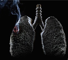 Cannot resist spreading this image from the Brazilian advertising agency
  The UCSF people are also experimenting with two other forms of online communication. To the right you can see
 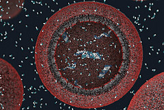 It turned out that this particular
  As you may have noticed, this blog has a crush on
 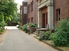 Yesterday morning, before our session on art and science, I took a walk through the beautiful old